
Muscle Fiber (Cell):
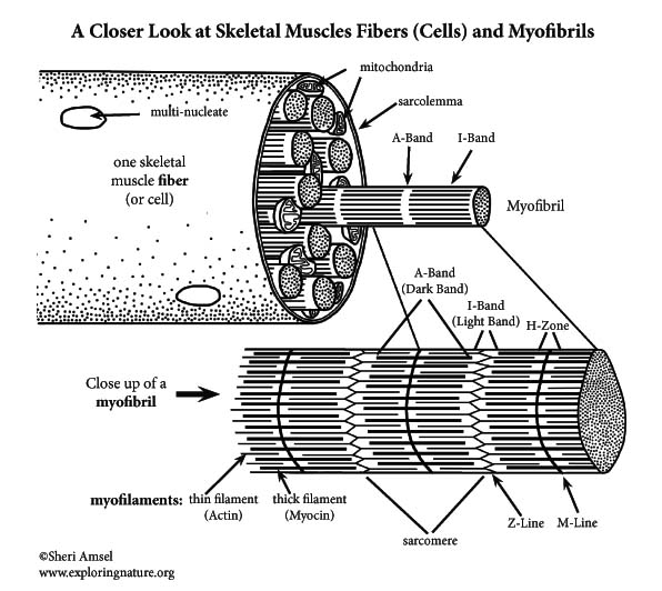
Each muscle fiber (cell) has tiny myofibrils running parallel in the cell. Myofibrils are the contractile elements of muscle cells.
When you look at a myofibril, you can see dark and light bands. The dark bands are A-Bands. The light bands are I-Bands. These are the visible striations you see in skeletal muscle.
This banding pattern of the myofibril is from protein filaments called myofilaments. The thick filaments extend the length of the A-Band. The thin filaments extend across the I-Band partway into the A-Band.
Relaxed Muscle Features:
In a relaxed muscle, inside the A-band there can be seen a light stripe called the H-Zone, which is lighter because the thick and thin filaments don’t overlap there.
The M-Line in the center of each H-Zone is where the thick filaments connect.
Down the center of the I-band there is a line called the Z-Line. This is the point of attachment of the thin filaments.
The region of the myofibril, between two Z-Lines is called a sarcomere. This is the smallest contractile unit of a muscle cell. Each myofibril is made up of chains of sarcomeres.
Myofibril Close Up:
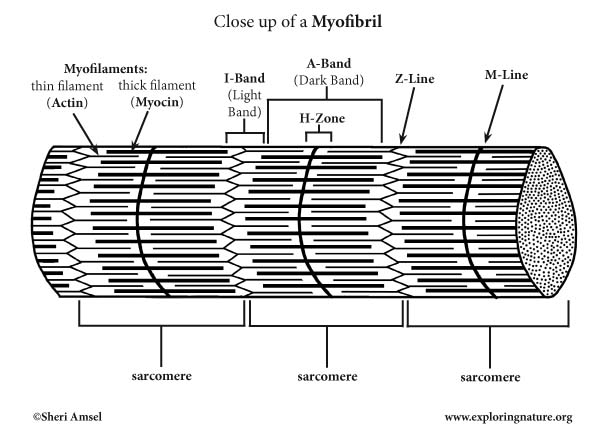
In a relaxed muscle, inside the A-band there can be seen a light stripe called the H-Zone, which is lighter because the thick and thin filaments don’t overlap there.
The M-Line in the center of each H-Zone is where the thick filaments connect.
Down the center of the I-band there is a line called the Z-Line. This is the point of attachment of the thin filaments.
The region of the myofibril, between two Z-Lines is called a sarcomere. Each myofibril is made up of chains of sarcomeres.
Test your knowledge of Myofibril Anatomy with the Myofibril Labeling page.

