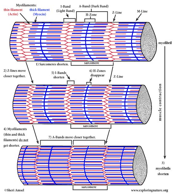

During a muscle contraction, every sarcomere will shorten (1).
This brings the Z-lines closer together (2).
The myofibrils shorten (3) too, as does the whole muscle cell.
Yet the myofilaments (the thin and thick filaments) do not get shorter (4). They slide by each other, overlapping as the Z-lines pull closer together.
The I-Bands shorten (5).
The H-Zones disappear (6).
The A-Bands move closer together (7).
How Muscle Contraction Works - Close Up
To understand how this happens, we will have to look closer to the myosin and actin contractile proteins.
When the nerve stimuli comes into the muscle cell to start a muscle contraction, the thick filaments’ myosin cross bridges (globular heads) grab onto an active site (1) on the thin filament (F-actin strands) adjacent to them and pull back (2) and release several times (think of a cranking ratchet: attach-pull-detach-reset).
This pulls the thin filament in toward the center of the sarcomere (3), shortening it over all. This reaction is happening at the same instant throughout the entire muscle (which received the stimuli), so the muscle contracts.
Muscle contractions (myosin cross bridge attachment) need calcium, which is released into the cell by the SR when the stimulus to contract occurs.

