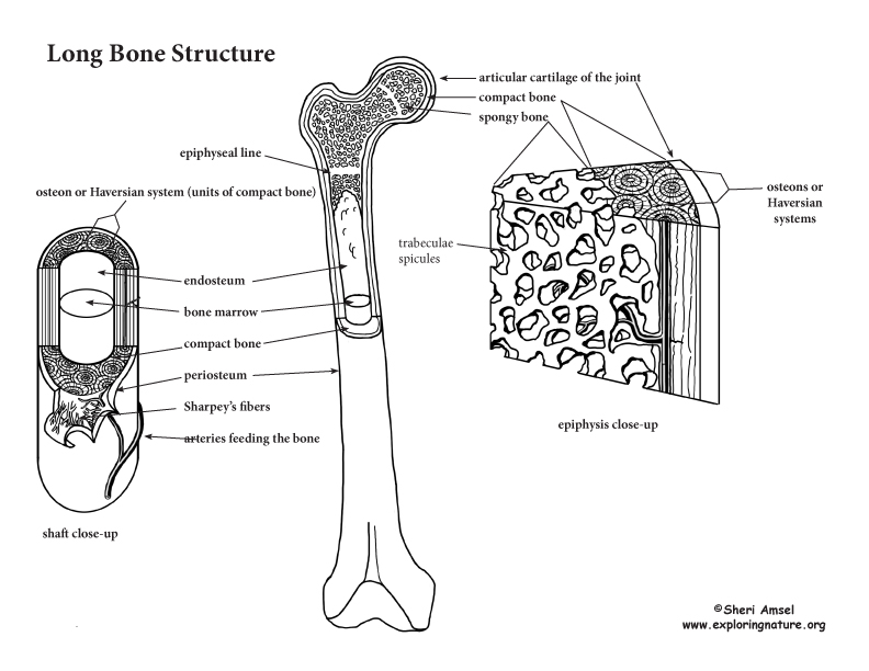

The long bones have a long, central shaft that enlarges at the ends into epiphysis. The long bones in the legs are the femur, tibia, and fibula. The long bones of the arms are the radius and ulna. Both the feet and hands have long bones in the digits – the phalanges. The adult femur is the largest bone in the body at about 2 feet long and is hollow to make it lighter. It is very strong to support the body’s weight, made up mostly of compact bone and some inner spongy bone (described below).
On the outside of living bone lies a layer of periosteum. The periosteum is the connective tissue covering that supplies blood vessels and nerves to bone. It is attached to the bone by Sharpies fibers. The inner side of the periosteum is lines by a layer of osteoblasts – cells that secrete the boney matrix (lay down more bone).
Inside the bone shaft there is a layer called endosteum. On the inner side surface of the endosteum of compact bone there is a layer of osteoclasts – cells that break down bone.
In adults, the central cavity of the long bones store yellow marrow. In children, the central cavity houses red marrow, which forms blood cells. Red marrow can be found in adult bones, but only in the epiphysis of spongy bone cavities.
Long bones are made up of a long, straight, central shaft with enlarged ends. The enlarged ends articulate (connect) with other bones. Instead of periosteum, the ends, called the epiphyseal surface, are covered by smooth hyaline cartilage. This “articular cartilage” creates a friction-free surface for joint movement.
In growing children, there is an epiphyseal plate toward the end of the long bones made up of hyaline cartilage for longitudinal growth. The epiphyseal plate transitions in adulthood to the epiphyseal line.
As bones grow, osteoblasts lay down more bone on the outside, while osteoclasts break down bone on the inside. This increases the diameter of the whole bone without decreasing the thickness of the compact bone walls. The hardness of bone is due to inorganic calcium salts in the ground substance (connective tissue) of the bone. The flexibility of bone is due to the collagen fibers.
Let’s compare compact bone to spongy bone on microscopic level:
Spongy bone is made up of trabeculae spicules. The trabeculae form a network of spaces filled with bone marrow. They act as struts to resist stress.
Compact bone is made up of osteons or Haversian systems. Each osteon is an elongated cylinder running up and down the bone – much like a weight bearing pillar. The osteon is laid down in layers or rings (like the rings of a tree) called lamella. Each osteon is 5-6 layers thick. The center of each osteon has a central canal or Haversian Canal that has blood vessels and nerve fibers.
For Assessment: Bone Anatomy Labeling
LS1.A: Structure and Function
• All living things are made up of cells, which is the smallest unit that can be said to be alive. An organism may consist of one single cell (unicellular) or many different numbers and types of cells (multicellular). (MS-LS1-1)
• Within cells, special structures are responsible for particular functions, and the cell membrane forms the boundary that controls what enters and leaves the cell. (MS-LS1-2)
• In multicellular organisms, the body is a system of multiple interacting subsystems. These subsystems are groups of cells that work together to form tissues and organs that are specialized for particular body functions. (MS-LS1-3)
